m Bot: removing deprecated anatomy infobox parameters (Task 11) |
m Dating maintenance tags: {{Merge}} |
||
| (31 intermediate revisions by 25 users not shown) | |||
| Line 1: | Line 1: | ||
{{short description|Carries tears from the lacrimal sac of the eye into the nasal cavity.}} |
|||
{{merge|nasolacrimal canal|date=March 2024}} |
|||
{{Infobox anatomy |
{{Infobox anatomy |
||
| Name = Nasolacrimal duct |
| Name = Nasolacrimal duct |
||
| Latin = |
| Latin = ductus nasolacrimalis |
||
| Greek = |
| Greek = |
||
| Image = Gray896.png |
| Image = Gray896.png |
||
| Line 8: | Line 10: | ||
| Image2 = Gray1199.png |
| Image2 = Gray1199.png |
||
| Caption2 = Outline of bones of face, showing position of air sinuses. |
| Caption2 = Outline of bones of face, showing position of air sinuses. |
||
| ImageMap = |
|||
| MapCaption = |
|||
| Precursor = |
| Precursor = |
||
| System = |
| System = |
||
| Line 16: | Line 16: | ||
| Nerve = |
| Nerve = |
||
| Lymph = |
| Lymph = |
||
| Code = |
|||
}} |
}} |
||
The '''nasolacrimal duct''' ( |
The '''nasolacrimal duct''' (also called the '''tear duct''') carries [[tears]] from the [[lacrimal sac]] of the [[Human eye|eye]] into the [[nasal cavity]].<ref name="Medscape 2020">{{Cite journal|date=2020-02-19|title=Nasolacrimal System Anatomy: Embryology, Puncta, Canaliculi|url=https://emedicine.medscape.com/article/835092-overview}}</ref><ref name="Herbert Janardhan Pandiri Cesta 2018 pp. 391–435">{{cite book | last1=Herbert | first1=Ronald A. | last2=Janardhan | first2=Kyathanahalli S. | last3=Pandiri | first3=Arun R. | last4=Cesta | first4=Mark F. | last5=Miller | first5=Rodney A. | title=Boorman's Pathology of the Rat | chapter=Nose, Larynx, and Trachea | publisher=Elsevier | year=2018 | isbn=978-0-12-391448-4 | doi=10.1016/b978-0-12-391448-4.00022-8 | pages=391–435 | s2cid=80517730 | quote=The paired nasolacrimal ducts carry lacrimal secretions from the eye to the nasal cavity, and originate as oval openings near the edge of the medial canthus of the eyelids. Initially the duct is small and circular, but in the middle portion, the diameter increases and the appearance is more oblong and saccular. The diameter again decreases before the duct enters the ventrolateral nasal vestibule medial to the root of the incisor tooth approximately 2 mm caudal to the nares.}}</ref> The duct begins in the [[eye socket]] between the [[maxilla]]ry and [[lacrimal bone]]s, from where it passes downwards and backwards. The opening of the nasolacrimal duct into the inferior [[nasal meatus]] of the nasal cavity is partially covered by a mucosal fold ([[Hasner's valve|valve of Hasner]] or ''plica lacrimalis'').<ref name="Merriam-Webster Medical Dictionary 2020">{{cite web | title=Medical Definition of PLICA LACRIMALIS | website=Merriam-Webster Medical Dictionary | date=2020-05-08 | url=https://www.merriam-webster.com/medical/plica+lacrimalis | access-date=2020-05-08 | quote=an imperfect valve at the opening of the nasolacrimal duct into the inferior meatus of the nose}}</ref> |
||
This is the reason the nose starts to run when a person is crying or has watery eyes from an allergy, and why one can sometimes taste eye drops. |
Excess tears flow through the nasolacrimal duct which drains into the inferior nasal meatus. This is the reason the [[Rhinorrhea|nose starts to run]] when a person is [[crying]] or has watery eyes from an allergy, and why one can sometimes taste [[eye drops]]. This is for the same reason when applying some eye drops it is often advised to close the nasolacrimal duct by pressing it with a finger to prevent the medicine from escaping the eye and having unwanted side effects elsewhere in the body as it will proceed through the canal to the nasal cavity. |
||
Like the lacrimal sac, the duct is lined by [[stratified columnar epithelium]] containing mucus-secreting [[goblet cells]], and is surrounded by connective tissue. |
Like the lacrimal sac, the duct is lined by [[stratified columnar epithelium]] containing mucus-secreting [[goblet cells]], and is surrounded by connective tissue. |
||
==Clinical significance== |
==Clinical significance== |
||
[[Nasolacrimal duct obstruction|Obstruction of the nasolacrimal duct]] may occur.<ref>[http://www.childrenshospital.org/views/june06/blocked_tear_ducts.html "Blocked tear ducts in infants"], ''Pediatric Views'', June 2006.</ref> This leads to the excess overflow of tears called ''[[epiphora (medical)|epiphora]]''. A congenital obstruction can cause cystic expansion of the duct and is called a [[dacryocystocele]] or ''Timo cyst''. Persons with dry eye conditions can be fitted with [[punctal plug]]s that seal the ducts to limit the amount of fluid drainage and retain moisture. |
[[Nasolacrimal duct obstruction|Obstruction of the nasolacrimal duct]] may occur.<ref name="CARREIRO 2009 pp. 13–97">{{cite book | last=CARREIRO | first=J | title=Pediatric Manual Medicine | url=https://archive.org/details/pediatricmanualm00doja | url-access=limited | chapter=Head and neck | publisher=Elsevier | year=2009 | isbn=978-0-443-10308-7 | doi=10.1016/b978-0-443-10308-7.00002-8 | pages=[https://archive.org/details/pediatricmanualm00doja/page/n12 13]–97 | quote=Dacryostenosis is a condition whereby the nasolacrimal duct is narrowed or blocked and the glandular secretions into the eye are prevented from draining properly. The secretions collect around the orifice of the duct and in the corner of the eye, where they thicken, resulting in a gooey, sticky substance that further clogs any drainage route.}}</ref><ref name="Stagner Jakobiec Eagle Charles 2018 pp. 648–685">{{cite book | last1=Stagner | first1=Anna M. | last2=Jakobiec | first2=Frederick A. | last3=Eagle | first3=Ralph C. | last4=Charles | first4=Norman C. | title=Diagnostic Pathology of Infectious Disease | chapter=Infections of the Eye and Its Adnexa | publisher=Elsevier | year=2018 | isbn=978-0-323-44585-6 | doi=10.1016/b978-0-323-44585-6.00021-7 | pages=648–685 | quote=Dacryocystitis is produced by obstruction of the nasolacrimal duct with resultant tear stasis. Clinically, patients display erythema and swelling of the lacrimal sac, creating a mass in the medial canthal area, centered below the medial canthal tendon.}}</ref><ref>[http://www.childrenshospital.org/views/june06/blocked_tear_ducts.html "Blocked tear ducts in infants"] {{Webarchive|url=https://web.archive.org/web/20120708152623/http://www.childrenshospital.org/views/june06/blocked_tear_ducts.html |date=2012-07-08 }}, ''Pediatric Views'', June 2006.</ref> This leads to the excess overflow of tears called ''[[epiphora (medical)|epiphora]]'' (chronic low-grade nasolacrimal duct occlusion).<ref name="Nerad Carter Alford 2008 pp. 131–137">{{cite book | last1=Nerad | first1=Jeffrey A. | last2=Carter | first2=Keith D. | last3=Alford | first3=Mark | title=Oculoplastic and Reconstructive Surgery | url=https://archive.org/details/rapiddiagnosisop00mdje | url-access=limited | chapter=Disorders of the Lacrimal System: Congenital Obstruction | publisher=Elsevier | year=2008 | isbn=978-0-323-05386-0 | doi=10.1016/b978-0-323-05386-0.50010-7 | pages=[https://archive.org/details/rapiddiagnosisop00mdje/page/n131 131]–137}}</ref> A congenital obstruction can cause cystic expansion of the duct and is called a [[dacryocystocele]] or ''Timo cyst''. Persons with dry eye conditions can be fitted with [[punctal plug]]s that seal the ducts to limit the amount of fluid drainage and retain moisture. |
||
During an ear infection, excess mucus may drain through the nasolacrimal duct in the opposite way tears drain. |
During an ear [[infection]], excess mucus may drain through the nasolacrimal duct in the opposite way tears drain. {{Citation needed|date=March 2021}} |
||
The canal containing the nasolacrimal duct is called the [[nasolacrimal canal]]. |
The canal containing the nasolacrimal duct is called the [[nasolacrimal canal]]. |
||
In humans, the tear ducts in males tend to be larger than the ones in females.<ref>[https://www.wsj.com/articles/SB10001424052748703922804576300903183512350 "Tears of Men and Women Are Different"], ''[[Wall Street Journal]]'', mirrored at [http://www.foxnews.com/health/2011/05/04/river-men-women-shed-different-tears/ "Why Men and Women Shed Different Tears"], [[Fox News]], May 5, 2011.</ref> |
In [[humans]], the tear ducts in males tend to be larger than the ones in females.<ref>[https://www.wsj.com/articles/SB10001424052748703922804576300903183512350 "Tears of Men and Women Are Different"], ''[[Wall Street Journal]]'', mirrored at [http://www.foxnews.com/health/2011/05/04/river-men-women-shed-different-tears/ "Why Men and Women Shed Different Tears"], [[Fox News]], May 5, 2011.</ref> |
||
==Additional |
==Additional image== |
||
<gallery> |
<gallery> |
||
Image:Gray196.png|Roof, floor, and lateral wall of left nasal cavity |
Image:Gray196.png|Roof, floor, and lateral wall of left nasal cavity |
||
</gallery> |
</gallery> |
||
==See also== |
==See also== |
||
{{Portal|Medicine|Anatomy}} |
|||
*[[Congenital lacrimal duct obstruction]] |
*[[Congenital lacrimal duct obstruction]] |
||
*[[Lacrimal apparatus]] |
*[[Lacrimal apparatus]] |
||
| Line 46: | Line 46: | ||
==References== |
==References== |
||
{{Reflist}} |
{{Reflist}} |
||
==External links== |
==External links== |
||
* {{SUNYAnatomyFigs|33|04|09}} |
* {{SUNYAnatomyFigs|33|04|09}} |
||
| Line 51: | Line 52: | ||
{{Accessory organs of the eye}} |
{{Accessory organs of the eye}} |
||
{{Authority control}} |
|||
{{DEFAULTSORT:Nasolacrimal Duct}} |
{{DEFAULTSORT:Nasolacrimal Duct}} |
||
Latest revision as of 18:31, 19 March 2024
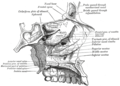
| Nasolacrimal duct | |
|---|---|
 The lacrimal apparatus. Right side. | |
 Outline of bones of face, showing position of air sinuses. | |
| Details | |
| Identifiers | |
| Latin | ductus nasolacrimalis |
| MeSH | D009301 |
| TA98 | A15.2.07.070 |
| TA2 | 6859 |
| FMA | 9703 |
| Anatomical terminology | |
The nasolacrimal duct (also called the tear duct) carries tears from the lacrimal sac of the eye into the nasal cavity.[1][2] The duct begins in the eye socket between the maxillary and lacrimal bones, from where it passes downwards and backwards. The opening of the nasolacrimal duct into the inferior nasal meatus of the nasal cavity is partially covered by a mucosal fold (valve of Hasner or plica lacrimalis).[3]
Excess tears flow through the nasolacrimal duct which drains into the inferior nasal meatus. This is the reason the nose starts to run when a person is crying or has watery eyes from an allergy, and why one can sometimes taste eye drops. This is for the same reason when applying some eye drops it is often advised to close the nasolacrimal duct by pressing it with a finger to prevent the medicine from escaping the eye and having unwanted side effects elsewhere in the body as it will proceed through the canal to the nasal cavity.
Like the lacrimal sac, the duct is lined by stratified columnar epithelium containing mucus-secreting goblet cells, and is surrounded by connective tissue.
Clinical significance
Obstruction of the nasolacrimal duct may occur.[4][5][6] This leads to the excess overflow of tears called epiphora (chronic low-grade nasolacrimal duct occlusion).[7] A congenital obstruction can cause cystic expansion of the duct and is called a dacryocystocele or Timo cyst. Persons with dry eye conditions can be fitted with punctal plugs that seal the ducts to limit the amount of fluid drainage and retain moisture.
During an ear infection, excess mucus may drain through the nasolacrimal duct in the opposite way tears drain. [citation needed]
The canal containing the nasolacrimal duct is called the nasolacrimal canal.
In humans, the tear ducts in males tend to be larger than the ones in females.[8]
Additional image
-
 Roof, floor, and lateral wall of left nasal cavity

See also
- Congenital lacrimal duct obstruction
- Lacrimal apparatus
- Nasolacrimal duct cyst
- Nasolacrimal duct obstruction
References
- ^ "Nasolacrimal System Anatomy: Embryology, Puncta, Canaliculi". 2020-02-19.
{{cite journal}}: Cite journal requires|journal=(help) - ^ Herbert, Ronald A.; Janardhan, Kyathanahalli S.; Pandiri, Arun R.; Cesta, Mark F.; Miller, Rodney A. (2018). "Nose, Larynx, and Trachea". Boorman's Pathology of the Rat. Elsevier. pp. 391–435. doi:10.1016/b978-0-12-391448-4.00022-8. ISBN 978-0-12-391448-4. S2CID 80517730.
The paired nasolacrimal ducts carry lacrimal secretions from the eye to the nasal cavity, and originate as oval openings near the edge of the medial canthus of the eyelids. Initially the duct is small and circular, but in the middle portion, the diameter increases and the appearance is more oblong and saccular. The diameter again decreases before the duct enters the ventrolateral nasal vestibule medial to the root of the incisor tooth approximately 2 mm caudal to the nares.
- ^ "Medical Definition of PLICA LACRIMALIS". Merriam-Webster Medical Dictionary. 2020-05-08. Retrieved 2020-05-08.
an imperfect valve at the opening of the nasolacrimal duct into the inferior meatus of the nose
- ^ CARREIRO, J (2009). "Head and neck". Pediatric Manual Medicine. Elsevier. pp. 13–97. doi:10.1016/b978-0-443-10308-7.00002-8. ISBN 978-0-443-10308-7.
Dacryostenosis is a condition whereby the nasolacrimal duct is narrowed or blocked and the glandular secretions into the eye are prevented from draining properly. The secretions collect around the orifice of the duct and in the corner of the eye, where they thicken, resulting in a gooey, sticky substance that further clogs any drainage route.
- ^ Stagner, Anna M.; Jakobiec, Frederick A.; Eagle, Ralph C.; Charles, Norman C. (2018). "Infections of the Eye and Its Adnexa". Diagnostic Pathology of Infectious Disease. Elsevier. pp. 648–685. doi:10.1016/b978-0-323-44585-6.00021-7. ISBN 978-0-323-44585-6.
Dacryocystitis is produced by obstruction of the nasolacrimal duct with resultant tear stasis. Clinically, patients display erythema and swelling of the lacrimal sac, creating a mass in the medial canthal area, centered below the medial canthal tendon.
- ^ "Blocked tear ducts in infants" Archived 2012-07-08 at the Wayback Machine, Pediatric Views, June 2006.
- ^ Nerad, Jeffrey A.; Carter, Keith D.; Alford, Mark (2008). "Disorders of the Lacrimal System: Congenital Obstruction". Oculoplastic and Reconstructive Surgery. Elsevier. pp. 131–137. doi:10.1016/b978-0-323-05386-0.50010-7. ISBN 978-0-323-05386-0.
- ^ "Tears of Men and Women Are Different", Wall Street Journal, mirrored at "Why Men and Women Shed Different Tears", Fox News, May 5, 2011.
External links
- Anatomy figure: 33:04-09 at Human Anatomy Online, SUNY Downstate Medical Center
- lesson9 at The Anatomy Lesson by Wesley Norman (Georgetown University)