| Brachioradial muscle | |
|---|---|
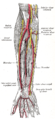
 Muscles of the upper extremity. | |
 Cross-section through the middle of the forearm. (Brachioradialis labeled at center left, sixth from the top.) | |
| Details | |
| Origin | Lateral supracondylar ridge of the humerus |
| Insertion | Distal radius (Radial styloid process) |
| Artery | radial recurrent artery |
| Nerve | radial nerve |
| Actions | Flexion of forearm |
| Identifiers | |
| Latin | musculus brachioradialis |
| TA98 | A04.6.02.039 |
| TA2 | 2496 |
| FMA | 38485 |
| Anatomical terms of muscle | |
Brachioradialis is a muscle of the forearm that acts to flex the forearm at the elbow. It is also capable of both pronation and supination, depending on the position of the forearm. It is attached to the distal radius and the lateral supracondylar ridge of the humerus.
Action
The brachioradialis flexes the forearm at the elbow. When the forearm is pronated, the brachioradialis tends to supinate as it flexes. In a supinated position, it tends to pronate as it flexes.
The brachioradialis is a stronger elbow flexor when the forearm is in a midposition between supination and pronation at the radioulnar joint. When pronated, the brachioradialis is more active during elbow flexion since the biceps brachii is in a mechanical disadvantage.
Innervation
Despite the bulk of the muscle body being visible from the anterior aspect of the forearm, the brachioradialis is a posterior compartment muscle and consequently is innervated by the radial nerve. Of the muscles that receive innervation from the radial nerve, it is one of only four that receive input directly from the radial nerve. The other three are the triceps, anconeus, and extensor carpi radialis longus. (All other posterior compartment muscles that receive radial innervation are supplied by the deep branch of the radial nerve.)
Appearance
If a person half-pronates their arm, to make a fist as if holding a handled vessel of beer, then puts their fist under a table or desk and tries to flex at the elbow, the brachioradialis will stand out of the forearm, visible under the skin.
Additional images
-
 Front of the left forearm. Superficial muscles.
 -
 Posterior surface of the forearm. Superficial muscles.
 -
 The radial and ulnar arteries.
 -
 Ulnar and radial arteries. Deep view.

External links
- Template:MuscleLoyola
- Illustration: brachioradialis from The Department of Radiology at the University of Washington
- Anatomy figure: 07:01-09 at Human Anatomy Online, SUNY Downstate Medical Center - "Transverse section through the left arm just proximal to the elbow."
- Anatomy figure: 07:03-07 at Human Anatomy Online, SUNY Downstate Medical Center - "Superficial muscles of the anterior (flexor) compartment of the left forearm."
- Anatomy figure: 09:02-02 at Human Anatomy Online, SUNY Downstate Medical Center - "Superficial muscles of the posterior (extensor) compartment of the left forearm."
- lesson5musofpostforearm at The Anatomy Lesson by Wesley Norman (Georgetown University)
- Anatomy image: Elbow5 at the College of Medicine at SUNY Upstate Medical University